Poster Presentation Annual Meetings of the Endocrine Society of Australia and Society for Reproductive Biology and Australia and New Zealand Bone and Mineral Society 2016
Oestradiol depletion in premenopausal women with non-metastatic breast cancer is associated with severely deteriorated cortical and trabecular microstructure (#335)
Introduction Treatment of premenopausal women with breast cancer using ovarian suppression (OS) and aromatase inhibition (AI) causes more rapid and complete oestradiol depletion than natural menopause. Oestrogen deficiency increases remodelling rate, prolongs osteoclast lifespan, and shortens osteoblast life span.1 Consequently, each of the many more remodelling events remove more bone more rapidly. We therefore hypothesised that the remodelling imbalance produces severe microstructural deterioration while the rapid remodelling reduces matrix mineral density (MMD) of the reduced matrix volume.
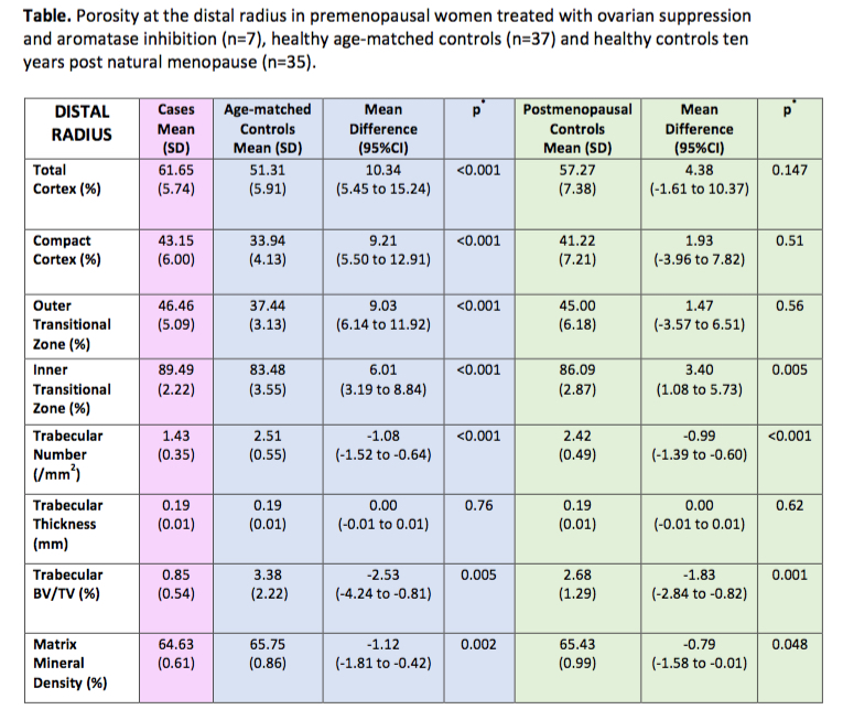
Methods At this early stage we have recruited 7 premenopausal women with breast cancer (mean age 45 years, range 38-51) treated with OS and AI for 38 months (range 11 – 118 months), 38 healthy age-matched premenopausal women and 38 women at least ten years post natural menopause (mean age 62 years, range 60-65). 6 cases had chemotherapy as part of their treatment. Women treated with tamoxifen for >6 months or anti-resorptives were excluded. Images of the distal radius and distal tibia were acquired using high-resolution peripheral quantitative computed tomography. Microstructure and MMD were quantified using StrAx1.0.2 Independent t-tests were used to compare morphology. Interim analysis was performed using SPSS v22.
Results Relative to premenopausal controls, cases had higher porosity of all cortical compartments, lower trabecular bone volume/total volume (BV/TV) due to fewer, not thinner trabeculae, and lower MMD (table). Despite being nearly two decades younger than women 10 years post natural menopause, cases had comparable or lower cortical porosity, lower BV/TV due to fewer, not thinner trabeculae, and lower MMD (table). Results at the tibia were similar (not shown).
Conclusions Severe and perhaps irreversible microstructural deterioration and the longevity of these women suggest there is a need to investigate the role of early intervention to preserve bone strength.
- 1. Manologas. Endocrine Reviews 2000; 21:115-37
- 2. Zebaze et al. Bone 2013; 54:8–20