Poster Presentation Annual Meetings of the Endocrine Society of Australia and Society for Reproductive Biology and Australia and New Zealand Bone and Mineral Society 2016
Synchrotron micro-CT time-lapsed imaging of the human femur microstructure under load (#277)
Introduction:
Time-lapsed micro-computed-tomography (micro-CT) with concomitant mechanical testing is increasingly used to study the bone deformation and fracture mechanism. However, previous femur studies were limited, imaging only small cores under load. We developed a protocol for time-lapsed synchrotron micro-CT imaging of entire human femoral epiphyses under load.
Methods:
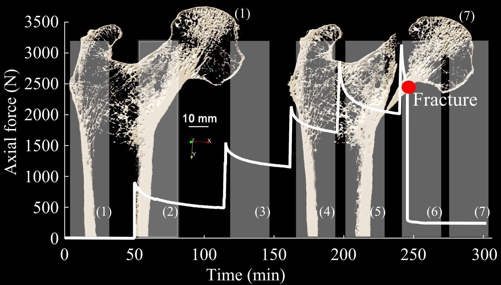
Twelve human femurs from elderly female donors (range: 56-91 years) were obtained. The fracture load was calculated using clinical CT images and finite-element modelling. A custom-made compression stage including an aluminum compression chamber, a 6-degree-of-freedom load cell and a screw-jack mechanism was manufactured. Samples were mounted inside the compressive stage, replicating a single-leg stance configuration. Micro-CT scans were performed at the Australian Synchrotron (31 μm/voxel, isotropic). One-fifth of the calculated fracture load was incrementally applied to the femur from the initial unloaded condition, with one micro-CT scan taken at each load step. At each step, the total volume scanned was 160 mm in diameter and 130 mm in height, scanning time 25 min. Four femurs were loaded to fracture, while 8 femurs were loaded non-destructively. The 6-component-force over time was recorded during the experiment.
Results:
Fractures were experimentally obtained in 5-6 load increments as predicted, with loads within the predicted range (1998-8636 N). The 2D and 3D images micro-CT images showed deformation and fracturing of the trabeculae and cortex. Sub-capital femoral neck fractures were obtained and were visible in the micro-CT images, consistent with observed patterns of clinical fractures (Figure 1).
Discussion:
Time-elapsed synchrotron micro-CT imaging of entire human femoral epiphyses with concomitant step-wise mechanical testing was successfully performed, at 31 μm voxel size. Clinically relevant fracture patterns were experimentally replicated and visible in the micro-CT images, together with the bone microarchitecture. Morphometric and micro-finite-element analyses are being undertaken, to investigate the contribution of the different microstructural compartments to withstand load.
H:\LAVORO_main_ALSOONLAPTOP\MEETINGS\ANZBMS\ANZBMS_2016_BRISBANE\SYNCHROTRON_ABSTRACT